Nuclear Medicine Imaging to support your Drug Development
Nuclear imaging plays a crucial role in preclinical research by offering a non-invasive and quantitative approach to visualize and analyze physiological and pathological processes in living organisms.
With 29 years of experience, Oncodesign Services is your partner for your preclinical studies combined with non-invasive imaging capabilities.
Introduction to nuclear imaging
Nuclear imaging is a medical imaging technique that uses radioactive tracers (radiotracers / radiopharmaceuticals) to visualize and assess the function of organs and tissues within the body. It involves the detection of gamma rays (SPECT imaging) or beta particles (PET imaging) emitted by these radioactive substances.
In the context of preclinical studies, nuclear imaging is commonly used to:
- Study physiological processes,
- Define pharmakokinetics and elimination routes of novel drugs
- Monitor disease progression,
- Evaluate the effectiveness of treatments in laboratory animals.
- Assess Dosimetry of novel radiopharmaceuticals for imaging and therapy
Radiotracers and nuclear imaging
Radiotracers used in preclinical nuclear imaging studies are often designed to target specific molecular pathways, receptors, or biological processes. This targeted approach allows researchers to gain insights into the molecular and cellular aspects of diseases and treatment responses.
Key technologies in nuclear imaging
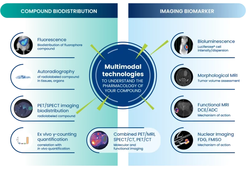
Nuclear imaging is often combined with other imaging techniques such as CT or MRI to provide complementary anatomical information. This fusion of imaging modalities enhances the overall understanding of the studied biological processes.
How Oncodesign Service supports your preclinical studies with nuclear imaging?
Oncodesign Services is a Contract Research Organization (CRO) specializing in nuclear imaging studies which can provide valuable support to biotech and pharmaceutical companies in preclinical studies.
Our experienced teams of scientists, radiochemists, and technicians in imaging techniques such as PET and SPECT contributing to the design and execution of effective nuclear imaging studies.
This expertise includes choosing appropriate radiotracers, optimizing imaging protocols, and determining the most suitable imaging modality for the research goals.


